
Ions in the Neurodegenerative Diseases (Part One)
Ions in the Neurodegenerative Diseases (Part One)

Most of us when talking about certain neurodegenerative diseases, prions is the first word that come up to our mind. The shrinkage of our neurons, the dementias, and the rest symptoms are not entertaining. The hardest part is almost there's no cure for those diseases.
Magnetite Pollution
Maher et al, 20161 reported a study about the magnetite pollution in the human brain. The team identified the abundant presence of magnetite nanoparticles (NPs) in the human brain. The NPs matched precisely the high-temperature magnetite nanospheres, formed by combustion and/or friction-derived heating. The NPs can enter the brain directly through the olfactory nerve. Nanoscale magnetite can respond to external magnetic fields and toxic to our brains. These will lead to the rise of reactive oxygen species (ROS) and linked to some neurodegenerative diseases such as Alzheimer's disease (AD). ROS can cause irreversible damage to DNA because ROS oxidize and modify some cellular components and prevent them from performing their original functions.
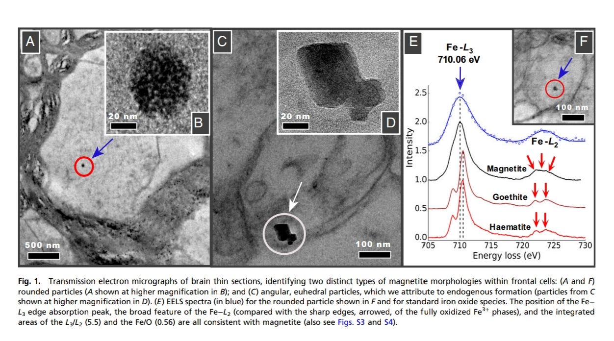
The team took 37 human brain samples, 29 from Mexico City and 8 from Manchester, UK. Researchers found magnetic contribution of ferromagnetic grains (<~20 nm), superparamagnetic (magnetically unstable) at room temperature. The NPs were not from biogenic ally formed magnetite, they were from the polluted air. There were Platinum (Pt), Nickel (Ni), Cobalt (Co), and possibly Copper (Cu) as additional presence in those brains.
The highest brain magnetite content is found in a 32 years old Mexico City resident. Many of the highly magnetic brain samples come from the older (>65 years old) Manchester cases, equivalent or higher magnetite concentrations were also displayed by young (<40 years old) Mexico City resident. The older cases had severe to moderate AD.
Aluminum (Al)
Al is highly reactive with carbon and oxygen, it is rapidly cleared by the kidney2. Al salts in vaccine adjuvants remind biologically available and accumulate in the nervous system. Al accumulation was found in the brain together with beta amyloid peptide accumulation. Al exposure increased the risk of AD by approximately 70%. In children with autism, Al level were found in the hair, blood, and urine. In the mice, aluminum hydroxide results in loss of long-term memory, increased anxiety, and neuronal death in the spinal cord and motor cortex. Many disease such as AD, amyotropic lateral sclerosis (ALS), autism spectrum disorders (ASDs), Guillain-Barre disease (GBD), multiple sclerosis (MS), and Gulf War syndrome (GWS) are associated with Al.

A study3 in 2020 reported that a higher brain aluminium content was found in sporadic Alzheimer’s disease (sAD), familial Alzheimer’s disease (fAD), ASD and multiple sclerosis (MS). Every atom of aluminium in human brain tissue must be accommodated as aluminium as Al3+(aq), is highly biologically reactive.
Ion Channels in Multiple Sclerosis
Ion channel dysfunction has been identified as a contributor to symptom development and neurodegeneration in MS4. Increased Na+ channel expression may contribute to neuronal energy insufficiency and a cascade of events that may ultimately lead to neurodegeneration and apoptosis.
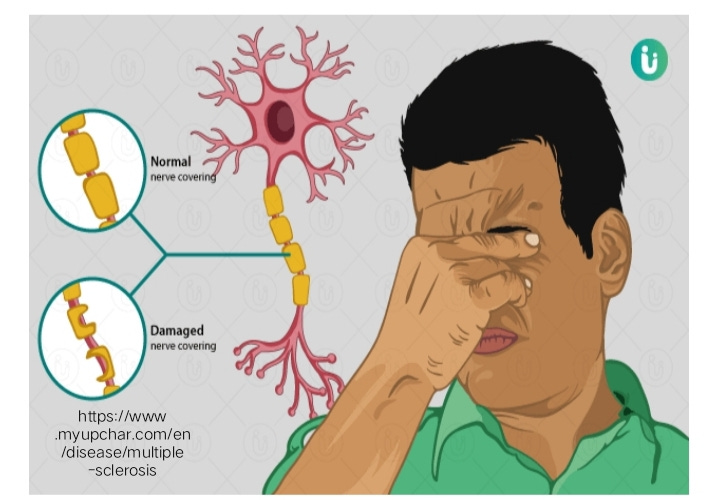
Na+ channel function have demonstrated pathological blockade of Na+ channels during an acute inflammatory attack. Pharmacological blockade of Na+ channels in animal models of MS demonstrated encouraging results. The process involved in demyelination, a characteristic event in MS pathology, may also induce complex structural changes mediated by K+ channels that may in turn hinder neural transmission.
Fig 1 https://www.pnas.org/content/113/39/10797
https://www.pnas.org/content/113/39/10797
https://pubmed.ncbi.nlm.nih.gov/27006759/
https://pubmed.ncbi.nlm.nih.gov/24202562/
https://www.eurekaselect.com/136233/article