Mitochondria and Mitochondria DNA in Some Diseases
Mitochondrial DNA as one of the inflammation & disease marker.
A better understanding of COVID-19 and it’s virus SARS-CoV-2 is needed. Many respected researchers have contributed through their proven researches and studies. Their greater responsibility is to the unfortunate COVID-19 patients and their families. Some of the researchers use the mitochondria as an approach to understand this virus and the disease.
Mitochondria are the place where our cell produces energy. They relate with energy production, innate immunity, ROS generation, apoptosis, Prasun, 20211. We can look for mitochondria in any cell of our body except the red blood cell (RBC). Mitochondria are located in and distributed throughout the cytoplasm, vary in number between different cell types. Each organelle is enclosed by a double membrane, with the inner one forming the characteristic folds known as cristae. Mutations causing mitochondrial dysfunction are often related to severe diseases, Protein Atlas2. In the Human Protein Atlas, we can see that 6% (1156 proteins) of all human proteins have been experimentally detected in the mitochondria. Mitochondrial proteins are mainly involved in cellular respiration and in mitochondrial organization, gene expression and metabolic processes. Mitochondria were abound in muscle tissue, especially human heart muscle tissues.
SARS-CoV-2 & Mitochondria
Wu et al., 20203 reported that majority of SARS-CoV-2 genomic and structural RNAs are targeted for mitochondrial matrix and nucleolus. SARS-CoV-2 RNA genome and all sgRNAs are enriched in the host mitochondrial matrix and nucleolus. The 5’ and 3’ viral untranslated regions possess the strongest and most distinct localization signals. Another researchers reported SARS-CoV-2 hijacked mitochondria machinery and DMV (double membrane vesicles), Singh et al.,20204. The hijacking may further compromise mitochondrial integrity, deteriorate mitochondrial function, and increase oxidative stress. An accumulation of damaged and dysfunctional mitochondria will lead to impaired MAVS (mitochondrial antiviral signaling) response, aggravates inflammation and cell death. SARS-CoV-2 prefers glycolysis and lactate accumulation. The damage of mitochondria impacts the decrease of oxidative phosphorylation as energy (ATP) source. Common mitochondrial supplements are Coenzyme Q10, vitamin B2, alpha lipoic acid, vitamin E, vitamin C, L-carnitine, L-creatine. N-acetylcysteine (NAC) is precursor of glutathione, an important intracellular antioxidant also can be used to prevent the mitochondria damage, Prasun, 2021.
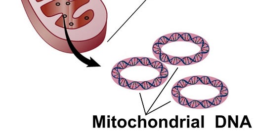
Mitochondrial DNA (mtDNA)
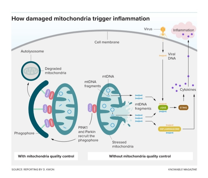
Mitochondria have their own DNA, mitochondrial DNA (mtDNA), member of a group of mitochondrial DAMPs (damaged-associated molecular patterns) released by injured or dying cells.
Circulating mtDNA levels were highly elevated in patients who eventually died or required ICU admission, intubation, vasopressor use, or renal replacement therapy, Scoozi et al., 20215. They reported that high circulating mtDNA was a potential early indicator for poor COVID-19 outcomes, suggesting mtDNA as investigational targets for COVID-19 therapy. The mtDNA are intrinsically inflammatory nucleic acids released by damaged solid organs. The release of mtDNA is often accompanied by the release of other mtDAMPs, induce inflammatory cytokine expression and the generation of reactive oxygen species (ROS) and the facilitation of neutrophil trafficking and activation.
The American Association for Clinical Chemistry (AACC) recommends mtDNA level as one of the markers of inflammation typically measured in COVID-19 patients, AACC6.
Animal studies and emerging clinical evidence indicate that exposure to chronic or severe stressors can alter mitochondrial DNA and mitochondrial function; this process may confer risk of psychiatric and other health conditions, Daniels et al.,20207.
Perez, 20178 reported that mtDNA mutations associated with pathogenic cases never lead (100% of the cases studied) to a reduction in long resonances (C+A)/(T+G) while hyper constraints super resonances decrease in 100% of the 250 cases of pathogenic mutations analyzed. This genomic optimality leads to an energetic over-functioning of the mitochondria of this pathogenic cell, which could explain the extreme pathogenicity and the disturbances observed in these pathogenic cells, especially in the cases of cancerous cells.
Abnormal expression of mtDNA in encoding proteins due to the defective of oxidative phosphorylation was observed. In cancer cells each cell contains many mitochondria with multiple copies of mtDNA, it is possible that wild-type and mutant mtDNA can co-exist in a state called heteroplasmy9.
Pictures:
2nd
https://www.liebertpub.com/doi/full/10.1089/dna.2020.6453
3rd https//knowablemagazine.org/article/mind/2021/could-mitochondria-be-key-healthy-brain?utm_campaign=K_newsletter-2021_06_20&utm_source=email&utm_medium=knowable-newsletter
https://www.liebertpub.com/doi/full/10.1089/dna.2020.6453
https://www.proteinatlas.org/humanproteome/cell/mitochondria
https://pubmed.ncbi.nlm.nih.gov/32511373/
https://journals.physiology.org/doi/full/10.1152/ajpcell.00224.2020?af=R
https://insight.jci.org/articles/view/143299
https://www.aacc.org/cln/articles/2021/march/mitochondrial-dna-is-early-marker-of-severe-covid-19-illness
https://www.annualreviews.org/doi/10.1146/annurev-clinpsy-082719-104030
https://www.researchgate.net/publication/318366048_Sapiens_Mitochondrial_DNA_Genome_Circular_Long_Range_Numerical_Meta_Structures_are_Highly_Correlated_with_Cancers_and_Genetic_Diseases_mtDNA_Mutations
https://www.nature.com/articles/1209604


